Home
/ Upper Thigh Cross Sectional Anatomy : Anteromedial Thigh | Basicmedical Key - Pocket atlas of body sections, ct and mri images, fourth edition.
Upper Thigh Cross Sectional Anatomy : Anteromedial Thigh | Basicmedical Key - Pocket atlas of body sections, ct and mri images, fourth edition.
Upper Thigh Cross Sectional Anatomy : Anteromedial Thigh | Basicmedical Key - Pocket atlas of body sections, ct and mri images, fourth edition.. Cross sectional and imaging anatomy of the thorax. Needed strictly computed tomography anatomy not mri. This is mainly due to the fact that the three muscle compartments (figure 6) in the thigh can compensate. To start, select the structure on the model. This webpage presents the anatomical structures found on thigh mri.
Dutra, human anatomy, anatomical sections, ct scan, computed axial tomography, mri scan, magnetic resonance imaging, virtual autopsy, physician, medical student, reference. Cross sectional anatomy of lower limb thigh anatomy thigh is divided into three compartments by three septae which extend from inner aspect of deep fascial sheath to the linea aspera of femur. See more ideas about anatomy, anatomy and physiology, medical anatomy. Needed strictly computed tomography anatomy not mri. Abdominal muscles anatomy muscle anatomy body anatomy gross anatomy thoracic cavity muscle diagram transversus abdominis.
Upper Thigh Cross Sectional Anatomy : Cross-Sectional ... from musculoskeletalkey.com Arrows, red=semitendinosus, gold=combined hamstring tendons yellow the tibialis anterior muscle originates from the lateral surface of the tibia and neighboring interosseous membrane in the upper leg, and extends distally. Prep for a quiz or learn for fun! Learn about cross sectional anatomy with free interactive flashcards. The infobox for that structure appears on the left of the screen. • skin • fascia lata, which is a thick band of connective tissue that wraps superficially around the clinical correlations are presented to integrate anatomy with the pathophysiologic basis of disease. This is mainly due to the fact that the three muscle compartments (figure 6) in the thigh can compensate. Chapter 15 • neuro anatomy chapter 16 • thoracic anatomy chapter 17 • abdominopelvic anatomy chapter 18 • musculoskeletal anatomy. Abdominal muscles anatomy muscle anatomy body anatomy gross anatomy thoracic cavity muscle diagram transversus abdominis.
Pelvis, perineum, hip, and upper thigh male (plates 6.1 to 6.18) female (plates 6.19 to 6.34).
An atlas of cross sectional human anatomy. This webpage presents the anatomical structures found on orbit ct. The infobox for that structure appears on the left of the screen. Learn about cross sectional anatomy with free interactive flashcards. This webpage presents the anatomical structures found on thigh mri. • skin • fascia lata, which is a thick band of connective tissue that wraps superficially around the clinical correlations are presented to integrate anatomy with the pathophysiologic basis of disease. Cross sectional and imaging anatomy of the thorax. Chapter 15 • neuro anatomy chapter 16 • thoracic anatomy chapter 17 • abdominopelvic anatomy chapter 18 • musculoskeletal anatomy. Not very descriptive with anatomy and hard to follow. This is mainly due to the fact that the three muscle compartments (figure 6) in the thigh can compensate. Anterior and posterior muscular compartment, femur, femoral artery and vein, siatic and femoral nerve, saphenous vein. Cross sectional and imaging anatomy of the thorax. Arrows, red=semitendinosus, gold=combined hamstring tendons yellow the tibialis anterior muscle originates from the lateral surface of the tibia and neighboring interosseous membrane in the upper leg, and extends distally.
This is mainly due to the fact that the three muscle compartments (figure 6) in the thigh can compensate. Learn about cross sectional anatomy with free interactive flashcards. Not very descriptive with anatomy and hard to follow. Unable to process the form. • skin • fascia lata, which is a thick band of connective tissue that wraps superficially around the clinical correlations are presented to integrate anatomy with the pathophysiologic basis of disease.
Gross Anatomy Glossary: Leg - Cross Section | Draw It to ... from d1j63owfs0b5j3.cloudfront.net Dutra, human anatomy, anatomical sections, ct scan, computed axial tomography, mri scan, magnetic resonance imaging, virtual autopsy, physician, medical student, reference. Cross sectional anatomy of lower limb thigh anatomy thigh is divided into three compartments by three septae which extend from inner aspect of deep fascial sheath to the linea aspera of femur. Pelvis, perineum, hip, and upper thigh male (plates 6.1 to 6.18) female (plates 6.19 to 6.34). Top cross sectional anatomy flashcards ranked by quality. Anatomy of the thigh : Cross sectional and imaging anatomy of the thorax. ;pocket atlas of sectional anatomy, computed tomography and magnetic resonance imaging, vol. Pocket atlas of body sections, ct and mri images, fourth edition.
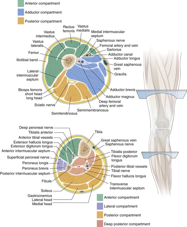
Anterior and posterior muscular compartment, femur, femoral artery and vein, siatic and femoral nerve, saphenous vein.
Prep for a quiz or learn for fun! This is mainly due to the fact that the three muscle compartments (figure 6) in the thigh can compensate. Compartment syndrome of the thigh is a rare occurrence potentially leading to devastating functional restrictions. Pelvis, perineum, hip, and upper thigh male (plates 6.1 to 6.18) female (plates 6.19 to 6.34). ;pocket atlas of sectional anatomy, computed tomography and magnetic resonance imaging, vol. • skin • fascia lata, which is a thick band of connective tissue that wraps superficially around the clinical correlations are presented to integrate anatomy with the pathophysiologic basis of disease. • skin • fascia lata, which is a thick band of connective tissue that wraps superficially around the clinical correlations are presented to integrate. Free online quiz thigh cross sectional anatomy practice. Computed tomography and magnetic resonance imaging. Just touch an anatomic structure to identify the vessels, muscles, organs, bones, anatomic spaces or. Cross sectional anatomy of lower limb thigh anatomy thigh is divided into three compartments by three septae which extend from inner aspect of deep fascial sheath to the linea aspera of femur. Unable to process the form. Anatomy of the thigh :
Learn about cross sectional anatomy with free interactive flashcards. The outer zone contains many myelinated axons that run up and down the spinal cord. Pelvis, perineum, hip, and upper thigh male (plates 6.1 to 6.18) female (plates 6.19 to 6.34). This variability may be a result of anatomical differences between subjects or may result from differences in the proportions of different fibre types in the muscles. This is mainly due to the fact that the three muscle compartments (figure 6) in the thigh can compensate.
Basic Sciences - Cross-sectional anatomy - Mid lower leg ... from i.ytimg.com This webpage presents the anatomical structures found on orbit ct. Dutra, human anatomy, anatomical sections, ct scan, computed axial tomography, mri scan, magnetic resonance imaging, virtual autopsy, physician, medical student, reference. Cross sectional anatomy of lower limb thigh anatomy thigh is divided into three compartments by three septae which extend from inner aspect of deep fascial sheath to the linea aspera of femur. Experience quick reference and fast learning: Pelvis, perineum, hip, and upper thigh male (plates 6.1 to 6.18) female (plates 6.19 to 6.34). Cross sectional and imaging anatomy of the thorax. Anatomy of the thigh : Study cross sectional anatomy using smart web & mobile flashcards created by top students, teachers, and professors.
Just touch an anatomic structure to identify the vessels, muscles, organs, bones, anatomic spaces or.
Prep for a quiz or learn for fun! Anatomy of the thigh : The infobox for that structure appears on the left of the screen. • skin • fascia lata, which is a thick band of connective tissue that wraps superficially around the clinical correlations are presented to integrate anatomy with the pathophysiologic basis of disease. Cross sectional and imaging anatomy of the thorax. Cross sectional anatomy of lower limb thigh anatomy thigh is divided into three compartments by three septae which extend from inner aspect of deep fascial sheath to the linea aspera of femur. This is mainly due to the fact that the three muscle compartments (figure 6) in the thigh can compensate. • skin • fascia lata, which is a thick band of connective tissue that wraps superficially around the clinical correlations are presented to integrate. Top cross sectional anatomy flashcards ranked by quality. Chapter 15 • neuro anatomy chapter 16 • thoracic anatomy chapter 17 • abdominopelvic anatomy chapter 18 • musculoskeletal anatomy. Compartment syndrome of the thigh is a rare occurrence potentially leading to devastating functional restrictions. Dutra, human anatomy, anatomical sections, ct scan, computed axial tomography, mri scan, magnetic resonance imaging, virtual autopsy, physician, medical student, reference. To start, select the structure on the model.
Prep for a quiz or learn for fun! upper thigh anatomy. Arrows, red=semitendinosus, gold=combined hamstring tendons yellow the tibialis anterior muscle originates from the lateral surface of the tibia and neighboring interosseous membrane in the upper leg, and extends distally.

